X-Ray and Computed Tomography laboratory
The Gips-Schüle Chair for Sustainable Systems Engineering runs a X-Ray CT laboratory with state-of-the-art equipment.
Besides a a high resolution industrial CT, a custom-built CT dedicated to advanced in-situ tomography is available.
industrial CT
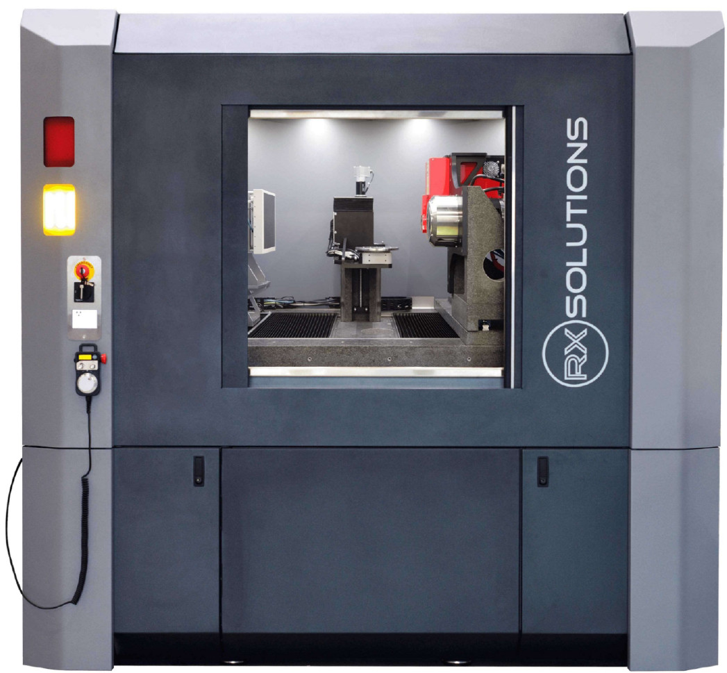
The EasyTom 150/160 CT system from RX Solutions is a versatile X-ray computed tomography system.
With a flexible combination of high-performance X-ray tubes (150 micro or 160 kV nano), it offers high image quality at resolutions better than one micrometer. Special features of the system include the choice of detector between a flat panel with 124 µm pixel pitch and a CCD with 9 µm pixel pitch, mechanical precision positioning of the sample sample, automatic scanning processes and a variety of measurement protocols, including e.g. laminography.
It is suitable for analyzing a wide range of materials and components, from small electronic electronic components, additively manufactured materials and larger industrial components. components. In-situ tensile and compression tests are possible in this system using a Deben CT5000 loadcell. Specimens with a diameter of between 5 to 20 mm can be subjected to forces up to 5 kN.
custom-built high-resolution dedicated in-situ CT
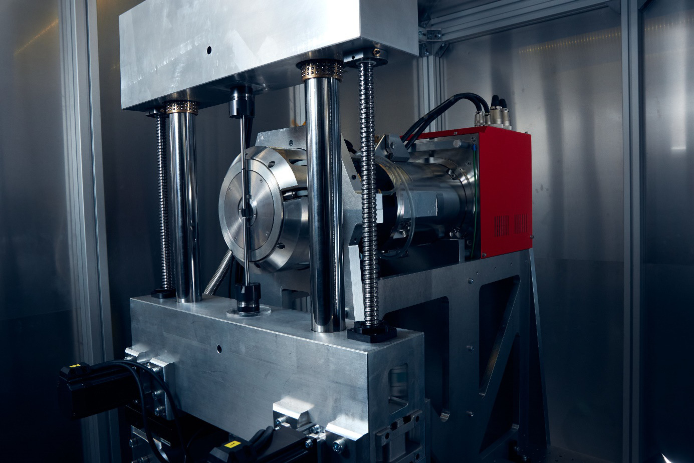
A system with an identical 160 kV nano-focus X-ray tube was developed and put into operation in collaboration with the Freiburg Materials Research Institute (FMF, Prof. Dr. Michael Fiederle). This system has an open load frame designed for this purpose. Compared to the Deben load frame, this enables a higher magnification factor and therefore a higher resolution for scans under load. Other advantages of the open system include improved accessibility and flexibility in sample placement as well as the ability to simulate various load scenarios under realistic conditions. A range of detectors including a conventional CMOS flat panel and photon-counting energy-sensitive techologies are available.
 |
| Figure 1: CT System „EasyTom 150/150“ from RX Solutions. |
 |
| Figure 2: custom-built dedicated high-resolution in-situ CT. The image shows the X-Ray source and the loading frame frame, which is capable of applying forces up to 10 kN. |
 |
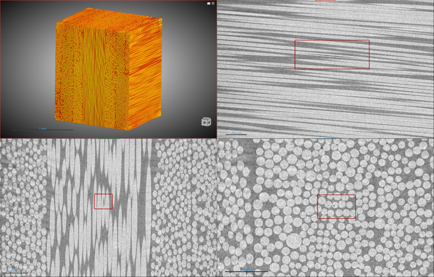
| Figure 3: CT image of a Basalt-fribre reinforced polymer. Voxel size is 800 nm. |
 |
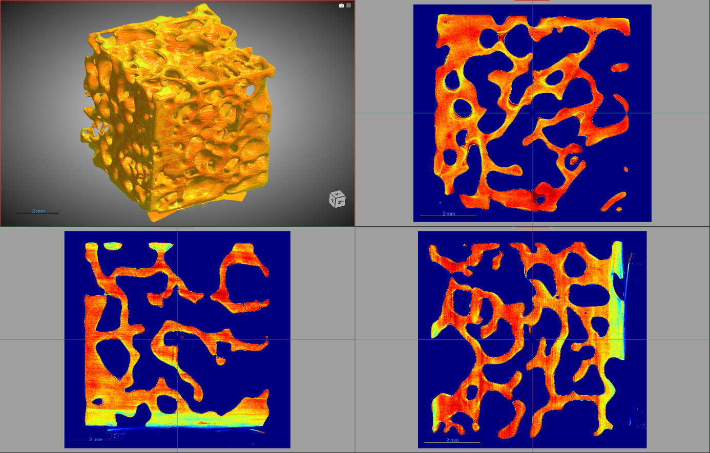
| Figure 4: CT reconstruction of a bone graft substitute in rendered 3D view and multiplanar sectional view Voxel size: 7 µm. |
associated publications:
- Plappert, D.; Schütz, M.; Ganzenmüller, G.C.; Fischer, F.; Campos, M.; Procz, S.; Fiederle, M.; Hiermaier, S. An Open-Frame Loading Stage for High-Resolution X-Ray CT. Instruments 2024, 8, 52. https://doi.org/10.3390/instruments8040052
- Fischer, F.; Plappert, D.; Ganzenmüller, G.; Langkemper, R.; Heusinger-Hess, V.; Hiermaier, S. A Feasibility Study of In-Situ Damage Visualization in Basalt-Fiber Reinforced Polymers with Open-Source Digital Volume Correlation. Materials 2023, 16, 523. https://doi.org/10.3390/ma16020523


